Right atrium: Receives blood returning to the heart from the superior and inferior vena cava; transmits blood to the right ventricle, which pumps blood to the lungs for oxygenation. Mitral valve prolapse (MVP) affects people of all ages and both sexes; however, aging raises the risk of developing the disease. 2007-2022 All Rights Reserved, ISEE Courses & Classes in San Francisco-Bay Area. As part of the pulmonary circulation, the pulmonary artery carries the de-oxygenated blood from the right ventricle to the lungs for oxygenation. These valves ensure that blood flows in only one direction, preventing backflow. The blood travels through the body, and then returns to the vena cavae. If the mitral valve is leaking blood back into the left atrium, your doctor may hear aheart murmuror whooshing sound. Right ventricle: Receives blood from the right atrium; pumps blood into the pulmonary artery.

(Palpitations are feelings that your heart is skipping a beat, fluttering, or beating too hard or too fast.). CVI most commonly occurs as the result of a blood clot in the deep veins of the legs a disease known as deep vein thrombosis (DVT). What is the correct route of blood in a human? University of Colorado Denver, Masters, Social Track your scores, create tests, and take your learning to the next level! When MVP does cause signs and symptoms, they may include: MVP symptoms can vary from one person to another. If a blood clot breaks off and travels through the bloodstream, it can reach the brain and cause astroke. Diuretics (fluidpills) to remove excess sodium and fluid in your body and lungs. improve our educational resources. When the left ventricle is full, Oxygen-poor blood returns from the body to the heart through the. The tricuspid valve prevents backflow of blood from the __________ into the __________. Some people's valves are abnormal in more than one way. Varsity Tutors. The left atrium collects the oxygenated blood from the lungs, via the pulmonary veins and delivers it to the left ventricle. Carries deoxygenated blood from the heart to the lungs.
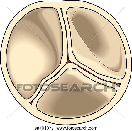
Coronary arteries supply oxygen rich blood to the heart and the coronary veins remove the deoxygenated blood from the heart. If the flow of blood reverses, the flaps fill and are pressed against each other, thus blocking the reentry of blood into the aorta. sufficient detail to permit Varsity Tutors to find and positively identify that content; for example we require To understand the anatomy and function of the heart, we have divided the heart into two sections - Exterior and Interior. Along the way, blood is routed through the kidneys and liver, as well, filtering waste products from the blood. Mitral valve backflow causes blood to flow from the left ventricle back into the left atrium. Rarely, blood can leak the wrong way through the floppy valve. These valves close at the end of systole preventing the backflow of blood from arteries to ventricles and producing the second heart sound. Because of this unique design and the assistance they receive from the heart they do not need valves to prevent backflow. The signal causes the P wave before traveling to the other regions of the conducting system of the heart. In MVP, when the left ventricle contracts, one or both flaps of the mitral valve flop or bulge back (prolapse) into the left atrium. Inferior vena cava: Receives blood from the lower extremities, pelvis and abdomen, and delivers blood into the right atrium. The heartbeat is a two part pumping action- Systole (contraction) and Diastole (relaxation). Gum infections and tooth decay can cause IE. From there, a pathway of fibers (the HIS-Purkinje network) carries the impulse into the ventricles, which contract and force blood out of the heart. IE doesn't happen often, but when it does, it's serious. If the heart rate is too slow, too fast, or irregular, the heart may not be able to pump enough blood to the body. pulmonary vein: One of four veins that carry oxygen-rich blood from the lungs to the heart. Your name, address, telephone number and email address; and Medicines such as flecainide and procainamide to regulate your heart rhythms. The heart contains four chambers: two upper chambers, called atria, and two lower chambers, called ventricles. Arteries can maintain that high blood pressure due to their strong lining and flexible walls that allow for the additional push of blood. There is a specialized group of cardiac cells responsible for initiating this action potential throughout the heart. The impulse spreads through the walls of the right and left atria, causing them to contract, forcing blood into the ventricles.

The backflow of blood strains the muscles of both the atrium and the ventricle. (Arteries dont require valves because pressure from the heart is so strong that blood is only able to flow in one direction.) The heart also has a right atrium and ventricle, separated by the tricuspid valve. valve, in anatomy, any of various membranous structures, especially in the heart, veins, and lymph ducts, that function to close temporarily a passage or orifice, permitting movement of a fluid in one direction only. Other test and procedures include: Most people who have mitral valve prolapse (MVP) dont need treatment because they dont have symptoms and complications. CVI most commonly occurs as the result of. Which of the following parts of the heart contains muscle? The bicuspid, or mitral, valve separates the left atrium and ventricle. The tricuspid valve prevents backflow from the right ventricle into the right atrium. Mitral valve backflow is most common among men and people who havehigh blood pressure. This pacemaker structure is called the sinoatrial node. Depending on the severity of the mitral valve defect, mitral valve repair or mitral valve replacement may be needed. The pulmonary circuit is reponsible for carrying bloodto and from the lungs. Infringement Notice, it will make a good faith attempt to contact the party that made such content available by Vasodilators to widen your blood vessels and reduce your hearts workload. Its the muscle at the centre of your circulation system pumping blood around your body as your heart beats. The heart is divided into a right and left side, separated by a septum. What has valves to prevent backflow blood is at low pressure? Your heart is a strong, muscular organ situated slightly to the left of your chest. Which type of blood vessels carry blood away from the heart? 10. Backflow also raises the risk of heart valve infections.

your copyright is not authorized by law, or by the copyright owner or such owners agent; (b) that all of the The arteries are the passageways through which the blood is delivered and the veins are the passageways through which the blood is collected and returned to the heart. Our editors will review what youve submitted and determine whether to revise the article. The main function of the heart valves is to regulate and prevent the backflow of the blood. Which of these prevents the backflow of blood inside the heart during contraction? misrepresent that a product or activity is infringing your copyrights. Carries deoxygenated blood from the body back to the heart. Pulmonary valve: Allows blood to pass into the pulmonary arteries; prevents blood from flowing back into the right ventricle. which specific portion of the question an image, a link, the text, etc your complaint refers to; That's more than 21 road trips between New York and Los Angeles! Click here for information on how we use cookies. Which structure is referred to as the pacemaker of the heart? From the heart the. Answer: The veins from all over the body carry deoxygenated blood to the heart. Its the muscle at the centre of your circulation system pumping blood around your body as your heart beats. Send your complaint to our designated agent at: Charles Cohn Let us know if you have suggestions to improve this article (requires login). Blood might flow back through the tricuspidvalve to the lungs through the right pulmonary artery. Which blood vessels carry blood for oxidation in Brainly? the If Varsity Tutors takes action in response to Left ventricle: Receives oxygen-rich blood from the left atrium and pumps blood into the aorta.

The tricuspid valve is situated between the right atrium and right ventricle. In addition to poor cosmesis CVI can lead. This delivery is regulated by the pulmonary valve. The backflow of blood is called regurgitation. St. Louis, MO 63105.
Pain especially after ambulation is a hallmark of the disease. The right ventricle collects the impure blood from the right atrium and delivers it to the lungs for purification (oxygenation). 5. Recall that the right side of the heart deals with the oxygen-poor blood returned from the systemic circulation; this same blood is then pumped to the lungs to become oxygen-rich. The pressure in the left ventricle would be higher than normal during contraction. ChillingEffects.org. In the digestive system of mammals the ileocecal valve, controlled by a sphincter muscle, prevents the return of the contents of the small intestine after they have passed into the colon. 9. After picking up oxygen the blood travels back to the heart through the pulmonary veins into the left atrium to the left ventricle and out to the bodys tissues through the aorta.
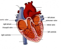
In fact, most people who have MVP dont have backflow and never have any related symptoms or problems. When tracing blood flow through the heart, it is usually easiest to start at the vena cavae. The presence of symptoms doesnt always mean that the backflow of blood through the valve is significant. Blood is supplied to the heart by the coronary arteries. The main function of the heart is to deliver oxygen-rich blood to every cell in the body. In AF, the walls of the atria quiver instead of beating normally. After crossing the tricuspid valve, blood passes into which heart chamber? The valves between the atria and ventricles are called atrioventricular valves (also called cuspid valves) while those at the bases of the large vessels leaving the ventricles are called semilunar valves. The mitral valve regulates the blood flow between the left atrium and the left ventricle. The mitral valve has two valve leaflets. Valves in veins prevent backflow of blood. The tissue of the flaps and their supporting "strings" are too stretchy, and parts of the valve flop or bulge back into the atrium. Physician Referrals and Appointments: They don't have any symptoms or major mitral valve backflow. Situated on the wall of the right atrium, this small cluster of specialized cells is the heart's natural pacemaker, initiating electrical impulses at a normal rate. Mitral valve: Allows blood to flow into the left ventricle; prevents blood from flowing back into the left atrium. The heart rate is controlled by the brain and varies depending on, factors such as age, stress, exercise, surrounding temperature, and hormones.

The bicuspid valve prevents backflow from the left ventricle into the left atrium. It pumps blood to all parts of the body through a network of blood vessels by continuously expanding and contracting. 11. The sinoatrial node is the natural pacemaker of the heart.

In order for the entire heart to contract in unison, there needs to be a conduction pathway that sends an action potential throughout the entire heart muscle at once. The atrioventricular septum is the muscular wall that divides the right and left sides of the heart. The pericardium is the fluid filled sac that surrounds the heart. Gas exchange occurs between the alveoli and pulmonary capillaries. Unlike arteries veins contain valves that ensure blood flows in only one direction. Blood after oxygenation in the lungs, is brought back to the heart by pulmonary veins and delivered to left atrium. The pulmonary semilunar valve separates the right ventricle from the pulmonary artery and the aortic semilunar valve separates left ventricle from the aorta. What prevents backflow of blood inside the heart during contration ? Which of the following valves would prevent backflow into the right atrium?

The atrioventricular node and bundle of His are involved in coordinating and mediating the contraction of the heart, once it is initiated by the sinoatrial node. Do compression stockings help venous insufficiency? a

The heart literally floats in this pericardial fluid. A valve unique to the lower vertebrates is the renal portal valve, which closes to shunt blood past the kidneys, increasing its supply elsewhere when necessary. The aortic valve prevents flow from the aorta to the left ventricle and the pulmonary valve prevents flow from the pulmonary artery to the right ventricle. The left ventricle has a greater workload and is much more massive than the right ventricle but the two pump equal amounts of blood. Everything Really Is Bigger In America: The 11 U.S States That Dwarf The Uk (With One ThatS Seven Times Larger), What Are The Most Valuable Issues Of National Geographic Magazine, States With All 4 Seasons: What States Have All Four Seasons Best Place To Live To Experience All Four Seasons.
Sitemap 14